Panic Disorder Risk Assessment Tool
This tool calculates your potential panic disorder vulnerability based on key neuroscience factors discussed in the article. Enter your current lifestyle factors to see how they affect your risk profile.
Calculate your risk
Enter your information above to see your personalized assessment.
Key Takeaways
- panic disorder is driven by over‑active threat circuits, especially the amygdala.
- Imbalances in serotonin, GABA and norepinephrine shape the intensity of panic attacks.
- The prefrontal cortex struggles to rein in the alarm system, creating a feedback loop.
- Stress hormones via the HPA axis and genetic factors set the stage for vulnerability.
- Therapies that target these brain mechanisms - CBT, exposure, SSRIs - can restore balance.
When you feel a sudden wave of dread that makes your heart pound and breath shallow, something concrete is happening inside your head. This article pulls back the curtain on the neuroscience of panic disorder, showing which brain structures light up, which chemicals go haywire, and how that knowledge guides modern treatment.
Panic disorder is a type of anxiety disorder characterized by recurrent, unexpected panic attacks and persistent concern about having more attacks affects roughly 2-3% of the population worldwide. While the experience feels purely emotional, decades of brain imaging and pharmacology reveal a cascade of physiological events.
How Panic Disorder Shows Up in the Brain
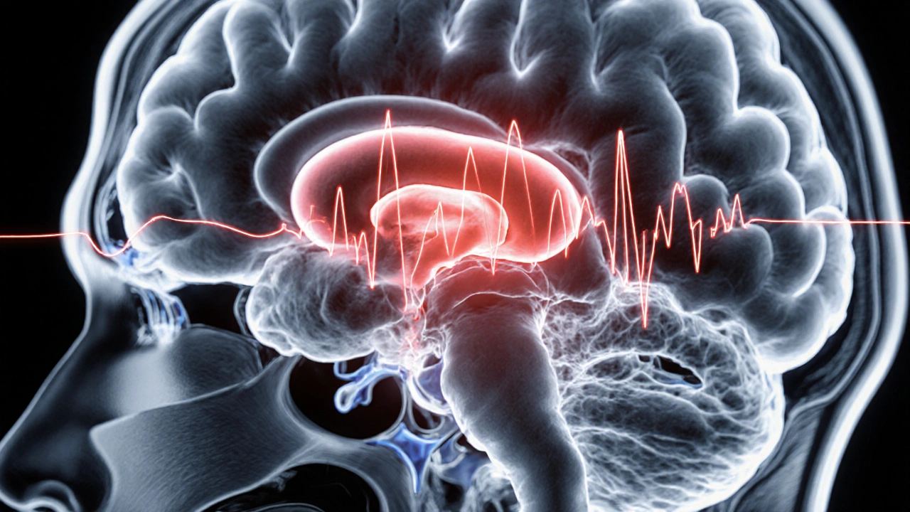
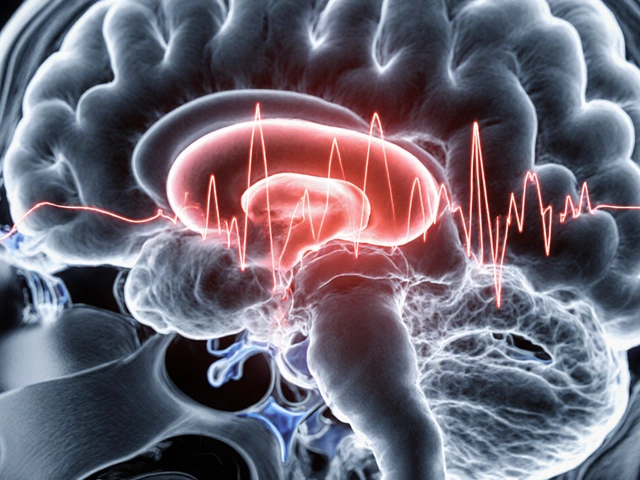
Functional MRI studies consistently show heightened activity in the brain’s threat detection network during a panic attack. The most reliable hotspots are the amygdala, the prefrontal cortex, and the hippocampus, all wired together by neurotransmitters and stress‑hormone pathways.
The Amygdala: Alarm System Gone Awry
Amygdala is an almond‑shaped cluster of nuclei deep within the temporal lobes that processes fear and emotional memory acts like an early‑warning radar. In people with panic disorder, the amygdala fires even without an external threat, sending a “danger” signal that triggers the autonomic nervous system. This explains the sudden surge of heart rate, sweating, and hyperventilation.
Neuroimaging shows the amygdala’s metabolic rate can be 30% higher during an induced panic episode compared to baseline, confirming its hyper‑reactivity.
Prefrontal Cortex: Lost Control
Prefrontal cortex is the front part of the brain responsible for decision‑making, impulse control, and moderating emotional responses normally reins in the amygdala. In panic disorder, functional connectivity between the prefrontal cortex and amygdala weakens, meaning the “brake” is less effective.
This weakening is most evident in the ventromedial prefrontal region, where activity drops by roughly 15-20% during attacks, undermining rational appraisal of the situation.
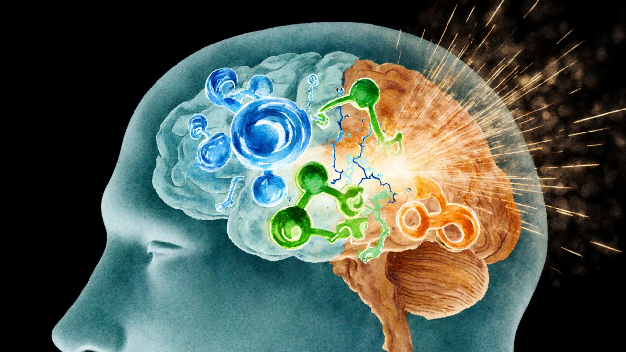
Neurochemical Imbalance: Serotonin, GABA, and Norepinephrine
Three neurotransmitters dominate the panic circuitry:
- Serotonin is a mood‑stabilizing neurotransmitter that modulates anxiety pathways helps keep the amygdala in check. Low serotonin transporter density correlates with higher panic frequency.
- GABA is the primary inhibitory neurotransmitter that calms neuronal firing deficiency removes the brain’s natural “off‑switch,” allowing unchecked excitation.
- Norepinephrine is a stress hormone neurotransmitter that ramps up alertness and heart rate spikes during panic, reinforcing the fight‑or‑flight response.
Medications that boost serotonin (SSRIs) or enhance GABA activity (benzodiazepines) directly target these chemical imbalances, which is why they can reduce attack severity.
The HPA Axis and Stress Hormones
HPA axis refers to the hypothalamic‑pituitary‑adrenal system that regulates stress hormones like cortisol acts as the body’s long‑term stress manager. Chronic hyper‑activation of the HPA axis raises baseline cortisol, which in turn sensitizes the amygdala.
Salivary cortisol studies show people with panic disorder often have a 10-15% higher morning cortisol level than healthy controls, suggesting a lingering stress imprint.
Genetics and Environmental Triggers
Family studies estimate that genetics account for about 40% of panic disorder risk. Specific gene variants-such as those in the serotonin transporter gene (5‑HTTLPR) that influences serotonin reuptake-are more common among patients.
However, genetics alone aren’t destiny. Traumatic experiences, chronic stress, or even caffeine over‑consumption can act as catalysts, tipping a vulnerable brain into the panic loop.
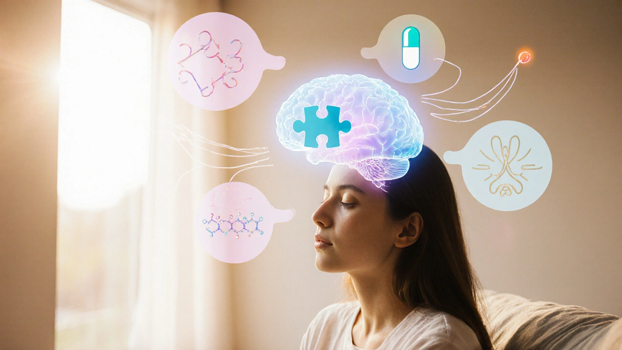
From Science to Treatment: Targeting the Brain
Understanding the neural circuitry guides three main therapeutic avenues:
- Cognitive Behavioral Therapy (CBT): Cognitive Behavioral Therapy is a structured, goal‑directed psychotherapy that challenges catastrophic thoughts and gradually exposes patients to feared sensations strengthens prefrontal control, restoring top‑down regulation of the amygdala.
- Medication: SSRIs elevate serotonin levels, while benzodiazepines boost GABA. Both dampen the hyper‑reactive threat circuitry.
- Mind‑body techniques: Breath training, mindfulness, and yoga reduce HPA‑axis output, lowering cortisol and norepinephrine spikes.
Combining CBT with an SSRI is often the most effective approach, as studies report remission rates around 60% versus 30% with medication alone.
Brain‑Region Comparison Table
| Region | Primary Role | Activity Change During Panic |
|---|---|---|
| Amygdala | Threat detection and fear conditioning | ↑ 30% (hyper‑active) |
| Prefrontal Cortex | Executive control, emotion regulation | ↓ 15-20% (reduced inhibition) |
| Hippocampus | Contextual memory, contextualizing fear | Variable; often ↓ volume over time |
| Insula | Interoceptive awareness (body sensations) | ↑ heightened perception of heartbeat |
Frequently Asked Questions
What triggers a panic attack in the brain?
A sudden surge of amygdala activity, often sparked by an internal cue (like a racing heartbeat) or an external stressor, overwhelms the prefrontal cortex. The resulting neurotransmitter cascade-high norepinephrine, low GABA, and insufficient serotonin-creates the classic physical symptoms.
Can genetics guarantee I’ll develop panic disorder?
No. Genetics raise susceptibility, but environment, lifestyle, and coping skills decide whether the disorder manifests. Most people with risk genes never experience panic attacks.
How fast do SSRIs work for panic disorder?
It typically takes 2-4weeks for serotonin levels to stabilize enough to reduce attack frequency, with full benefits often seen after 8-12weeks.
Is breathing technique enough to stop an attack?
Controlled breathing can blunt the physiological surge and signal the prefrontal cortex that the threat is manageable. It rarely ends an attack on its own but is a valuable adjunct to therapy.
What role does the HPA axis play in long‑term panic risk?
Chronically elevated cortisol from a hyper‑active HPA axis sensitizes the amygdala, making it fire more easily. Over time, this creates a feedback loop that sustains panic vulnerability.

Michelle Dela Merced
October 12 2025OMG this panic brain stuff is wild! 😱
Mark Conner
October 18 2025Listen, the U.S. has the best mental health research, and this article nails the science behind panic attacks.
Charu Gupta
October 23 2025The exposition delineates the neurobiological substrates of panic disorder with commendable precision, yet certain terminological inconsistencies warrant correction.
Carl Boel
October 29 2025From a neuroendocrine perspective, hyperactivation of the HPA axis constitutes a quintessential biomarker for acute panic episodes, underscoring the indispensability of cortisol assays.
Shuvam Roy
November 3 2025If you’re struggling with panic, prioritizing consistent sleep, moderate exercise, and CBT can rewire prefrontal inhibition pathways and lower HPA drive.
These lifestyle tweaks are backed by solid neuroscience and are easy to implement day‑to‑day.
Jane Grimm
November 9 2025While the interactive tool is aesthetically pleasing, its algorithmic oversimplification betrays a superficial grasp of the underlying neurochemistry.
Nora Russell
November 14 2025The author’s reductionist framework neglects the bidirectional circuitry between the amygdala and ventromedial prefrontal cortex, thereby rendering the discussion intellectually impoverished.
Craig Stephenson
November 20 2025I see your point, but for most readers the simplified model might actually help them take actionable steps.
Eve Perron
November 25 2025Understanding panic disorder through a neuro‑biological lens has profound implications for both treatment and stigma reduction.
First, the amygdala’s hyper‑reactivity serves as the alarm system that can trigger a cascade of physiological responses.
Second, the prefrontal cortex, when functioning optimally, acts as the brake that moderates this alarm.
When the brake fails, the hypothalamic‑pituitary‑adrenal (HPA) axis releases cortisol, amplifying the fear response.
Research shows that mindfulness‑based stress reduction can strengthen prefrontal regulation, thereby dampening HPA activation.
Moreover, regular aerobic exercise has been demonstrated to increase GABAergic inhibition, which further quiets the amygdala.
Pharmacologically, selective serotonin reuptake inhibitors (SSRIs) boost serotonergic tone, improving both mood and top‑down control.
Psychotherapy, particularly cognitive‑behavioral therapy (CBT), teaches patients to reinterpret catastrophic thoughts, effectively retraining neural pathways.
It’s also worth noting that genetics play a role; polymorphisms in the SLC6A4 gene influence serotonin transporter efficiency.
Environmental factors, such as chronic stress or traumatic events, can epigenetically modify gene expression, heightening vulnerability.
Therefore, a comprehensive treatment plan should address both biological and psychosocial dimensions.
In practice, clinicians can use the risk‑assessment tool to tailor interventions-high HPA scores may warrant early pharmacotherapy, while low prefrontal scores suggest a focus on CBT.
Patients should be encouraged to monitor sleep hygiene, as sleep deprivation exacerbates amygdala reactivity.
Finally, reducing caffeine intake can lower baseline arousal, making the nervous system less prone to over‑reaction.
By integrating lifestyle modifications with evidence‑based therapies, we can empower individuals to reclaim control over their panic responses.
Josephine Bonaparte
December 1 2025Great breakdown! Your points on sleep and caffeine really hit home – I’ve felt the difference myself when I cut back on evening coffee.
Meghan Cardwell
December 6 2025From a clinical standpoint, combining SSRIs with structured exposure therapy yields the highest remission rates for panic disorder, as meta‑analyses confirm.
stephen henson
December 12 2025Keep pushing forward – every small habit change adds up! 💪
Rhys Black
December 17 2025Honestly, the article’s attempt to simplify the neurocircuitry feels like a tragic oversell – we deserve a richer, more nuanced narrative.
Abhishek A Mishra
December 23 2025Appreciate the practical tips, especially the bit about regular walks – it’s simple but super effective.